EN
EN CN
CN
Foot & Ankle MSK Ultrasound: Core Podiatry Imaging for Plantar Fascia and Tendon Disorders
Podiatric care heavily relies on reliable musculoskeletal imaging to diagnose chronic foot and ankle pain that plagues athletes, office workers, and aging populations alike. Conventional X-rays only capture bony structures, failing to visualize soft tissue layers like ligaments, tendons, fascia, and subcutaneous edema—tissues responsible for over 70% of podiatric outpatient complaints. Foot ultrasound podiatry has become the gold standard bedside imaging tool, delivering real-time, radiation-free visualization to pinpoint subtle lesions without costly MRI scans or lengthy hospital appointments. For podiatrists, MSK ultrasound ankle workflows streamline triage, guide minimally invasive injections, and track rehabilitation progress for recurring foot pathologies, from mild plantar fascia tightness to degenerative Achilles tendinopathy.
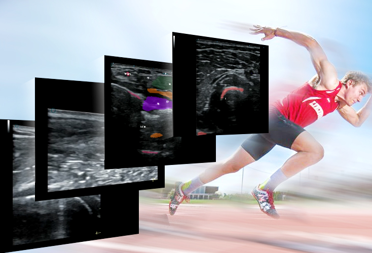
Plantar Fascia Ultrasound: Diagnosing and Grading Plantar Fasc11tis
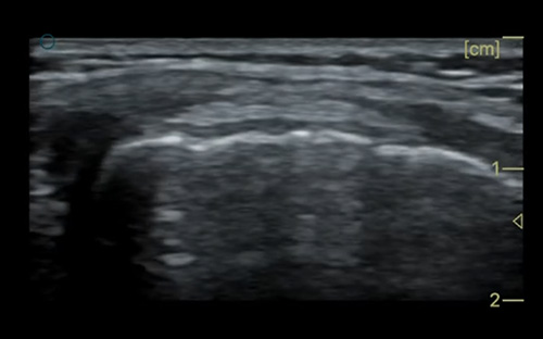
Plantar fasc11tis remains the most prevalent chronic heel pain condition, triggered by repetitive impact, flat foot biomechanics, obesity, or prolonged standing. Plantar fascia ultrasound allows clinicians to measure fascia thickness, detect fiber tears, and identify interstitial edema or calcific spurs buried within soft tissue—details invisible on plain radiographs. Normal plantar fascia measures less than 4mm at its calcaneal insertion; thickening above 5mm signals inflammatory degeneration, while hypoechoic splits indicate partial or full-thickness tears. Handheld ultrasound plantar fasc11tis diagnosis enables instant bedside grading: mild inflammatory thickening, moderate partial tearing, or severe complete rupture. Podiatrists use these quantitative measurements to design targeted shockwave therapy, orthotic interventions, or ultrasound-guided trigger point injections, eliminating guesswork from conservative treatment planning. Serial follow-up scans also track fascia healing over weeks, adjusting rehabilitation load to avoid re-injury.
Ankle Tendon Ultrasound: Evaluating Achilles Tendinopathy and Peroneal Lesions
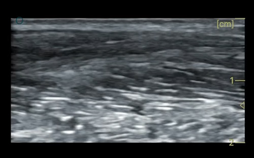
Achilles tendinopathy is a leading injury among runners, basketball players, and individuals with tight calf musculature, marked by tendon swelling, micro-tears, and degenerative mucoid changes. Portable ultrasound Achilles tendinopathy scans map tendon fiber architecture, distinguish reactive tendinosis from full tendon rupture, and quantify tendon width to gauge disease severity. Beyond the Achilles tendon, ankle tendon ultrasound assesses peroneal, tibialis posterior, and extensor tendon complexes, common sources of lateral ankle instability and midfoot pain after sprains. Color Doppler ultrasound further highlights neovascularization within damaged tendons—a key marker of active inflammation that dictates anti-inflammatory treatment protocols. Unlike MRI, ultrasound lets podiatrist dynamically move the patient’s foot during scanning, reproducing pain-provoking positions to link imaging findings directly to clinical symptoms for accurate differential diagnosis.
Ultrasound Foot Soft Tissue Evaluation for Complex Podiatric Presentations
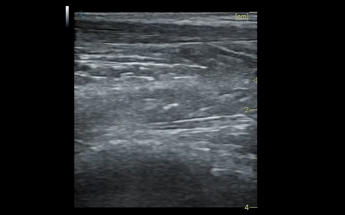
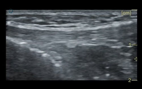
Ultrasound foot soft tissue evaluation extends far beyond tendons and fascia to cover a full spectrum of podiatric pathologies. Clinicians utilize linear transducer imaging to assess Morton’s neuromas between metatarsal heads, subcutaneous bursitis, plantar fibromas, and post-sprain soft tissue hematomas. For diabetic foot care, ultrasound detects hidden abscesses, tracking fluid collections beneath intact skin to prevent progressive ulceration and osteomyelitis. Dynamic scanning also evaluates ligamentous laxity in unstable ankles, measuring talar shift during inversion/eversion stress tests to guide bracing or reconstructive surgery referrals. This versatile imaging modality eliminates the need for delayed cross-sectional imaging for routine podiatric complaints, cutting patient wait times and enabling immediate, same-day treatment plans in private podiatry clinics and sports medicine facilities.
Clinical Workflow Advantages of Podiatry Ultrasound in Daily Practice
The widespread adoption of podiatry ultrasound stems from its unique practical benefits for foot and ankle specialists. First, it is entirely radiation-free, making it safe for repeated serial scans in pediatric patients, pregnant individuals, and long-term chronic pain sufferers. Second, real-time dynamic imaging allows clinicians to correlate anatomical abnormalities with patient-reported pain during active foot movement, a capability static MRI cannot match. Third, ultrasound-guided interventions—such as corticosteroid injections for plantar fasc11tis or barbotage for Achilles calcifications—improve precision and reduce off-target tissue damage compared to blind injection techniques. For community podiatry offices with limited imaging budgets, portable linear ultrasound systems remove reliance on external radiology centers, accelerating patient care and boosting clinic operational efficiency.
Conclusion
Musculoskeletal ultrasound has revolutionized modern podiatry, delivering accessible, precise soft tissue assessment for plantar fascia, ankle tendon, and general foot soft tissue disorders that dominate foot pain caseloads. Accurate grading of plantar fasc11tis, early detection of Achilles degenerative changes, and dynamic evaluation of foot biomechanics all depend on high-resolution foot ultrasound podiatry to guide evidence-based treatment. For podiatric clinics seeking a compact, cost-effective scanning tool to integrate daily MSK ankle assessments, PAU1A linear handheld ultrasound delivers crisp superficial soft tissue imaging, adjustable gain and Doppler modes, and multi-device wireless connectivity, perfectly suited for routine plantar fascia and tendon evaluations in busy foot care practices.