Tel/Whatsapp: +86 15005204265
Email: [email protected]
Tel/Whatsapp: +86 15005204265
Email: [email protected]
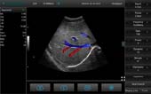
Color Mode Liver 3
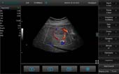
Color Mode Liver
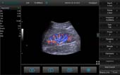
Color Mode Kidney 1
For more insights of lesions
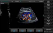
Color Mode Kidney 2
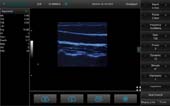
B Mode Carotid
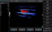
Color Mode Carotid
Color Mode Carotid1
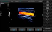
For more insights of lesions

Color Mode Thyroid3
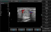
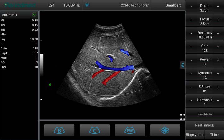
Convex Probe-Color Mode-Liver 3
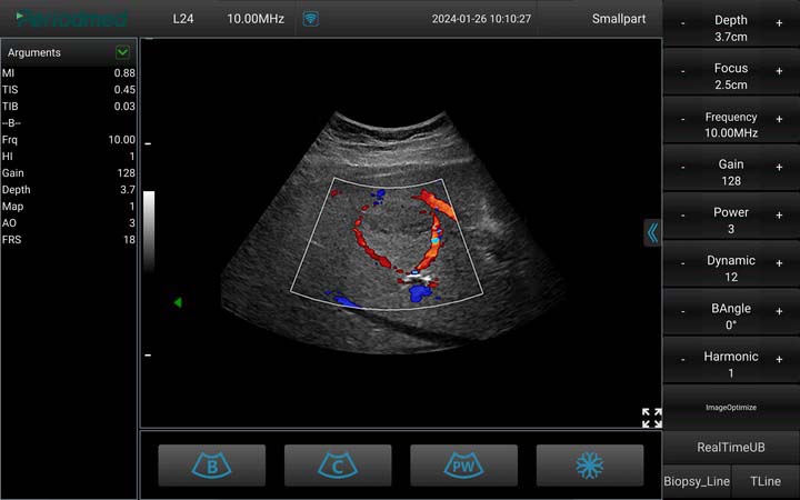
Convex Probe-Color Mode-Liver
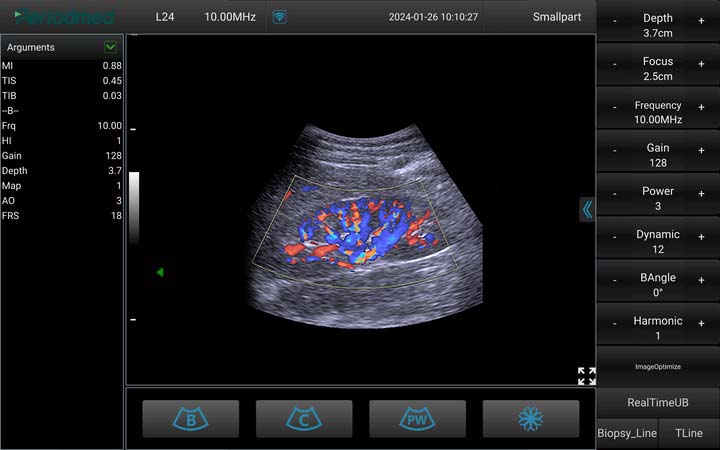
Convex Probe-Color Mode-Kidney 1
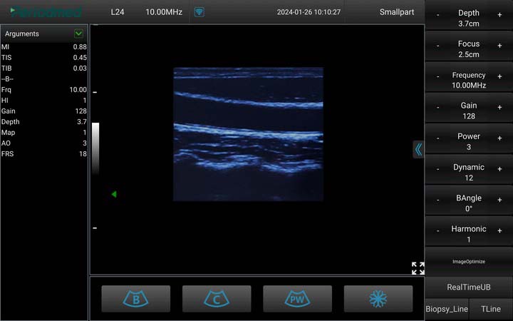
Linear Probe-B Mode-Carotid
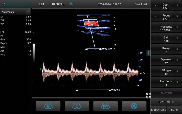
Linear Probe-PW Mode-Carotid
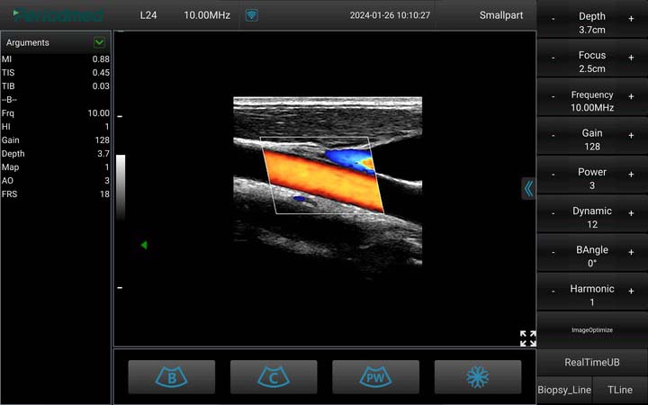
Linear Probe-Color Mode-Carotid3
Element: | 192array |
Gain Control: | 8 Segment TGC and overall gaincan be adjustable |
| Language Support: | English,Arabic, Chinese,French, German, Indonesian, Italian, Portuguese,Russian,Spanish |
Storage formats: | DICOM |
Image formats: | BMP, JPG |
Storage devices: | Hard drive image storage |
Application: | Generic,Abdomen,Obstetrics,Gynecology,Vascular,Small Parts, Urology, MSK,Cardiology |
Scanning Angle/length: | Convex array 60degrees,Linear array 40mm,Phased Array 90 degree |
| Frequency: | Convex array 2.5-6.0MHz, Linear array5.0-12.0 MHz, Phased array 2.5-6.0MHz |
| Measurement: | Length, Area,Speed,Heart rate,S/D, Velocity,Obstetric, Automatic blood flow |

Display Modes:B Mode,CF Mode, M Mode, PW Mode, Power Doppler Imag-
ing, Directional Power Doppler Imaging, Color M Mode,Nee-dle Enhance Mode
Image adjustment:Gain,Depth,Freg,Dynamic range,Focus,lmage enhance-ment,Biopsy,Automatic optimization,Spatial compound im-aging, Speckle reduction imaging,Color quantification, Virtual convex,Fine angle steer,Tissue harmonic imaging,Pulse in-version harmonic imaging