Tel/Whatsapp: +86 15005204265
Email: [email protected]
- Home
- Product
Ultrasound System
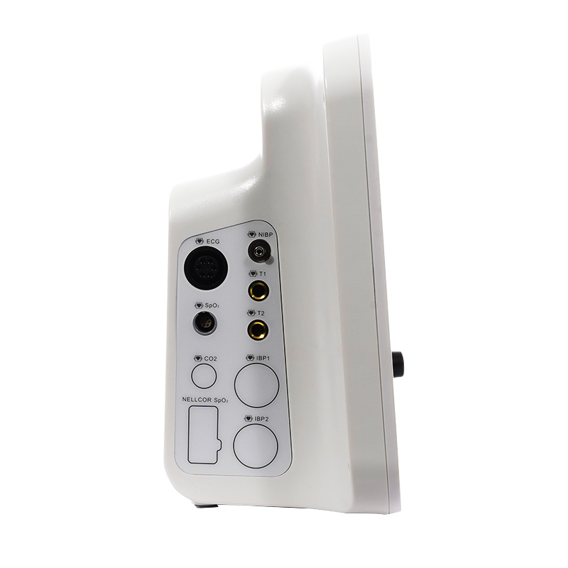
Life Information Monitoring System
Infusion & Syringe System
Solution
- About
- Media & Events
- Service & Support
- Contact Us
Tel/Whatsapp: +86 15005204265
Email: [email protected]

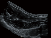
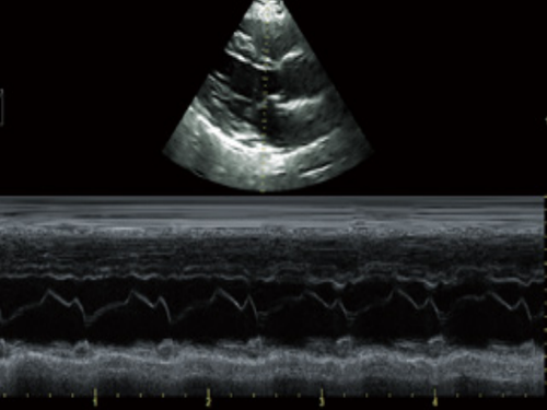
Cardiac B + M + CF
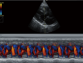
Cardiac M mode

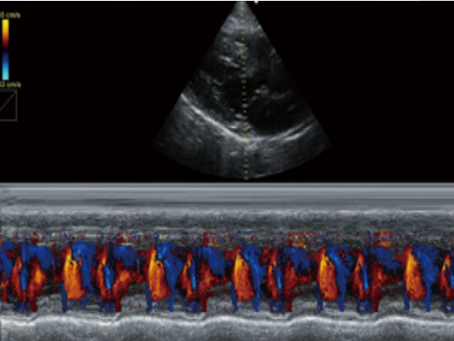
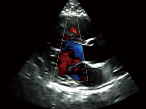
Cardiac CF mode

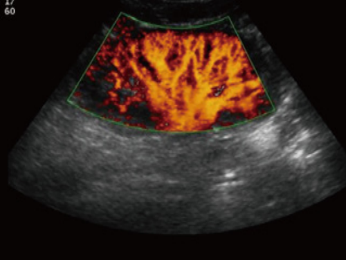
Color Liver image
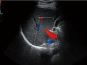
Kidney PDI mode

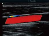
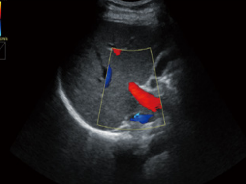
Carotid CF mode
Panorama image
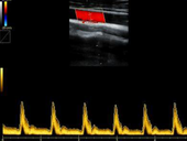
Carotid B + C + D mode
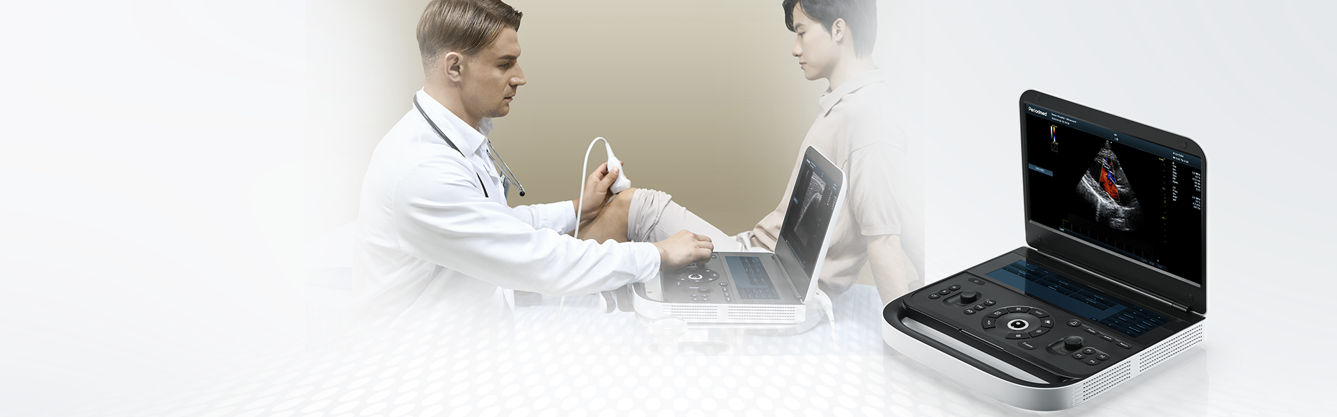
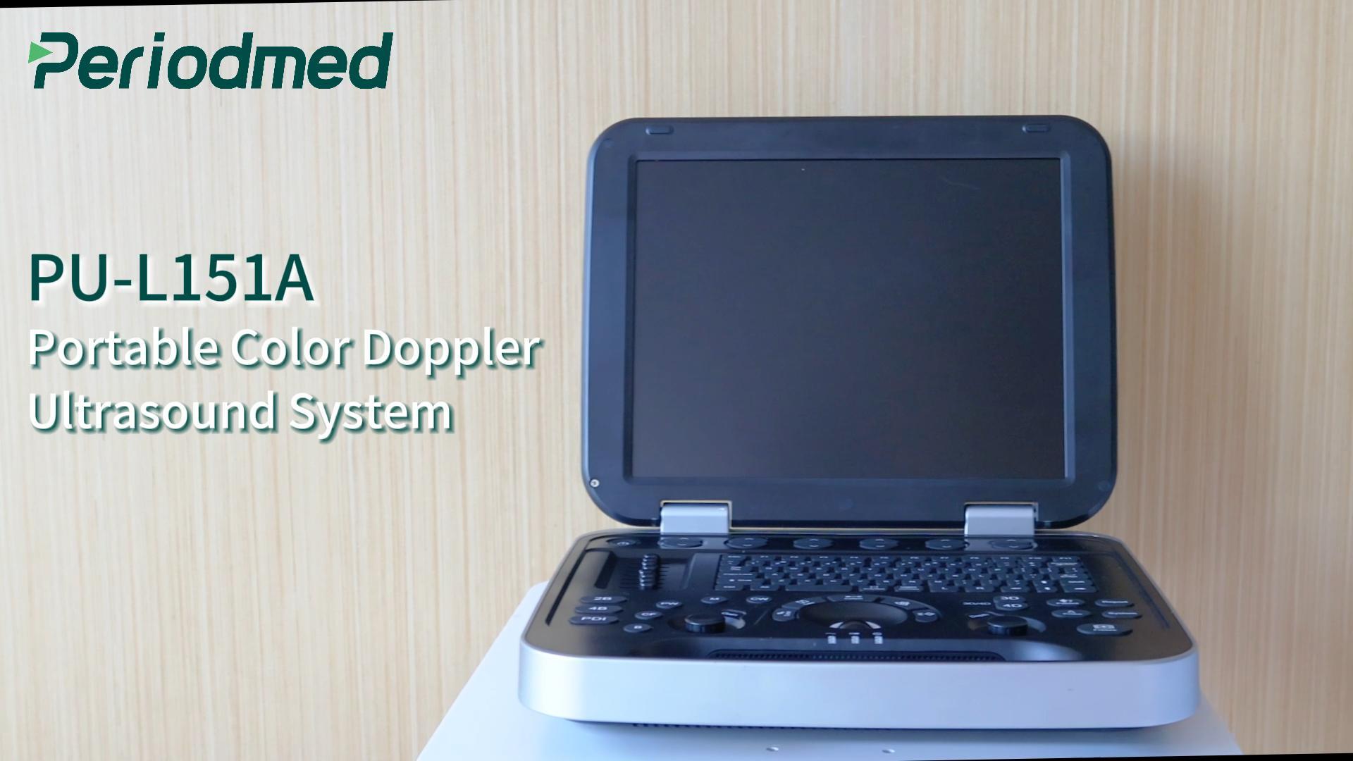
PU-L151A is an abdominal ultrasound imaging device for abdominal ultrasound using high-frequency sound waves to create images of internal organs like the liver, gallbladder, pancreas, and kidneys. It not only helps doctors to diagnose conditions such as gallstones, kidney stones, or tumors but also general applications.
Abdominal ultrasound imaging device works seamlessly for abdominal ultrasound procedure, during which a healthcare professional applies a gel to the abdomen and moves a transducer over the skin. Transducer sends sound waves that reflect off organs to form a sonogram (computer image). L151A can be used to diagnose or monitor conditions affecting the liver, gallbladder, kidneys, and major abdominal blood vessels.
Using adaptive signal processing technology, the echo signal of the undetermined area is analyzed by the unique data-only sensing method, which improves the resolution and uniformity of the image and easily obtains the high-definition heart image. Using the second harmonic and nonlinear fundamental wave imaging, the excellent signal-to-noise ratio image is obtained, which provides a more accurate basis for clinical judgment. Improved contrast resolution and enhanced visualization are achieved by using multiple scanned angles to create a single image.
Allowing adjustments to the scan line to gain better visibility of the needle, nerves and small vessels. Broadens the scope of spatial relationships by showing the entire anomaly and its relationship to adjacent structures on a single static image. Through detailed observation and analysis of the heart structure, doctors can more accurately judge the type and extent of the disease and develop a more appropriate treatment plan for patients.

| Application | |||
| Generic | Obstetrics | Gynecology | Abdomen |
| Vascular | Small Parts | Urology | MSK |
| Cardiology | |||
| Transducer Types | |||
| Convex probe | Linear probe | Micro Convex probe | Trans-vaginal probe |
| Volume probe | Trans-rectal probe | ||
| Standard Mode | |||
| B | 2B | 4B | PW |
| CF | M | PDI | Panorama |
| Trapezoid | Color M | DPDI | PIP |
| Anatomy M | |||
| System Architecture | |||
Physical Channels: 16 | Elements: 80 | Frequency Range: 2.0-22.0 MHz | Dynamic Range: ≥120dB |
| Beam forming : 4 | Hard Drive:128G | 15 Inches High Resolution LED Monitor | Resolution:1024*768 |
| Memory: 4G | |||
| Optional Mode | |||
| Wide view | 3D/4D mode | Needle enhance mode | Contrast enhance mode |


 EN
EN CN
CN