Tel/Whatsapp: +86 15005204265
Email: [email protected]
- Home
- Product
Ultrasound System
Life Information Monitoring System
Infusion & Syringe System
Solution
- About
- Media & Events
- Service & Support
- Contact Us
Tel/Whatsapp: +86 15005204265
Email: [email protected]
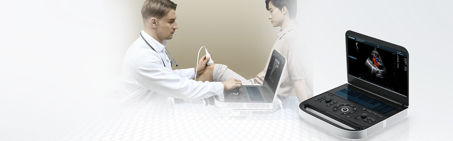
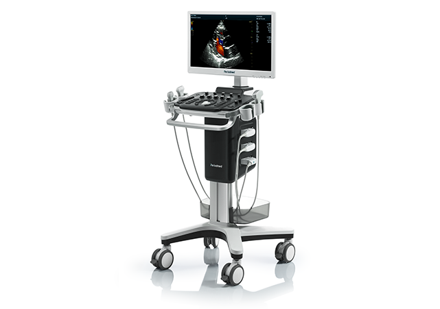
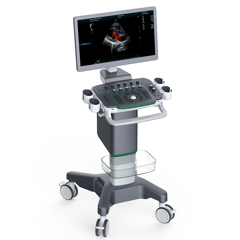
Transforming Primary Care Efficiency | Revo T1

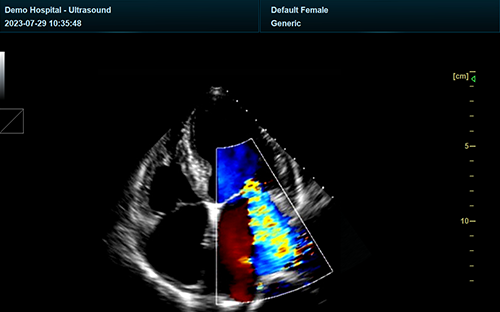
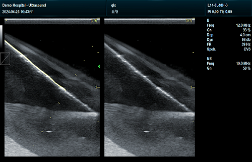
Revo T1 is a powerful cart-based needle enhancement ultrasound system with a goal to improve the needle's appearance on the screen. Needle enhancement ultrasound uses a combination of ultrasound beam steering and specialized algorithms to increase the visibility of a needle during ultrasound-guided procedures, resulting in a bright, clear line that aids in accurate needle placement for tasks like biopsies and nerve blocks.
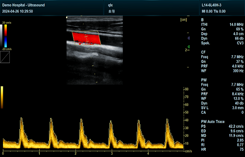
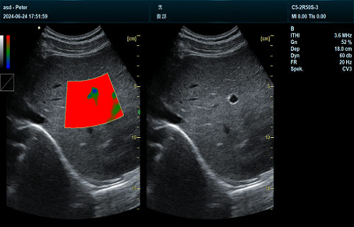
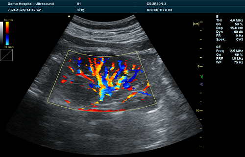
Powered by the latest holographic domain technology, Periodmed offers ultrasound images with extraordinary clarity for needle enhancement. The details representation and scanning fluidity guarantee excellent quality of ultrasound examinations across different applications including biopsies, treatment injections, anesthesia, and more.
The hardware design of Revo T1 needle enhancement ultrasound machine is engineered to refine examination workflows, positioning it as a highly efficient platform. Special care is taken during the manufacturing of this ultrasound system to incorporate environmentally friendly materials—panels included—thereby advancing Periodmed’s commitment to building a more sustainable world.
Revo T1 gains an edge from a point-and-shoot focused-domain optical imaging platform—powered by holographic domain technology. It smashes through the limits of traditional ultrasonic beam synthesis, delivering a more accurate basis for needle enhancement ultrasound image assessment. It also blends intelligent blue-light enhancement technology which automatically detects the needle, optimizing image quality for safer, precise, and efficient puncturing.
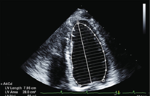
The extremely high imaging sensitivity and comfortable grip design make it easy to use, expanding the clinical usage scenarios. Single - Crystal transducers provide deeper penetration with needle enhancement ultrasound details; ComboWave transducers enhance imaging sensitivity; The core structure of the piezoelectric chip is used to obtain real - time scanning focusing imaging.
| Application | |||
| Generic | Obstetrics | Gynecology | Abdomen |
| Vascular | Small Parts | Urology | Thyroid |
| Cardiology | MSK | ||
| Image Formats | |||
| AVI | BMP | JPG | PNG |
| DICOM | |||
| Transducer Types | |||
| Convex probe | Linear probe | Phased array | Micro convex array |
| Volume array | Intracavity probe | ||
| Operating Mode | |||
| B | 2B | 4B | CW |
| M | CF | B/M | PW |
| PDI | Picture in Picture | Trapezoid | Panorama |
| 3D/4D(Optional) | Wide view(Optional) | Color M Mode | Anatomy M |
| TDI(Optional) | Needle Enhance Mode(Optional) | Contrast Enhance Mode(Optional) | |
| System Architecture | |||
| Elements: 64/128 | Frequency Range: 2.0-22.0 MHz | Channel:64 | Hard Drive:128G |
| Dynamic Range: ≥120db | 13.3 Inches touch screen | Resolution:1024*768 | Beam forming 4 |
| Memory:4G | 21 Inches High Resolution LED Monitor | ||
| Triplex | |||
| B+CF | PDI | DPDI+PW | |
| Languages | |||
| English | Arabic | French | Indonesian |
| Italian | Portuguese | Spanish | Chinese |
| German | Russian | ||


 EN
EN CN
CN