Tel/Whatsapp: +86 15005204265
Email: [email protected]
- Home
- Product
Ultrasound System
Life Information Monitoring System
Infusion & Syringe System
Solution
- About
- Media & Events
- Service & Support
- Contact Us
Tel/Whatsapp: +86 15005204265
Email: [email protected]
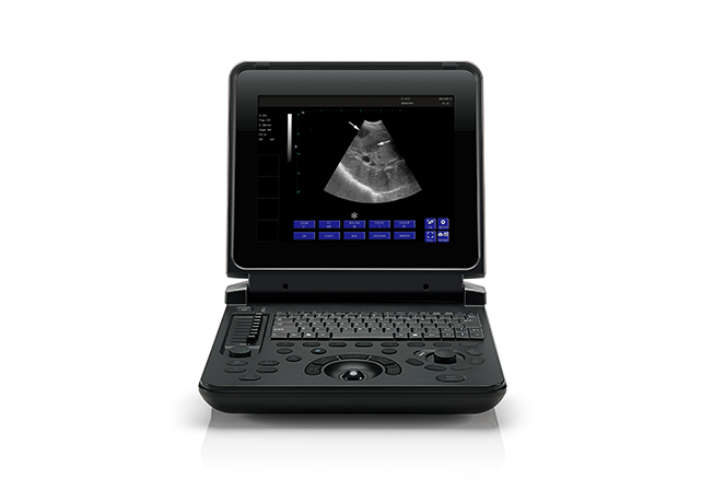
Black and White Ultrasound Machine | PU-DL121A

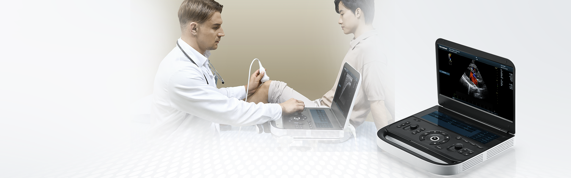
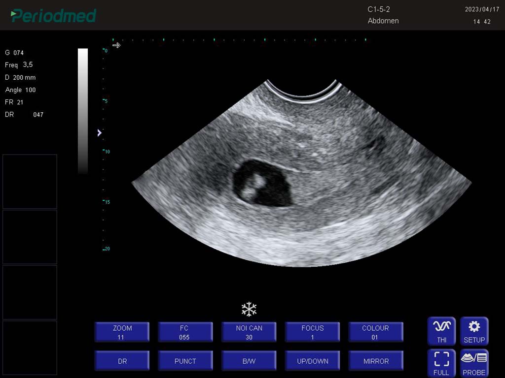
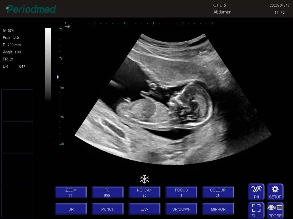
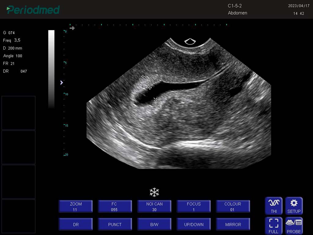
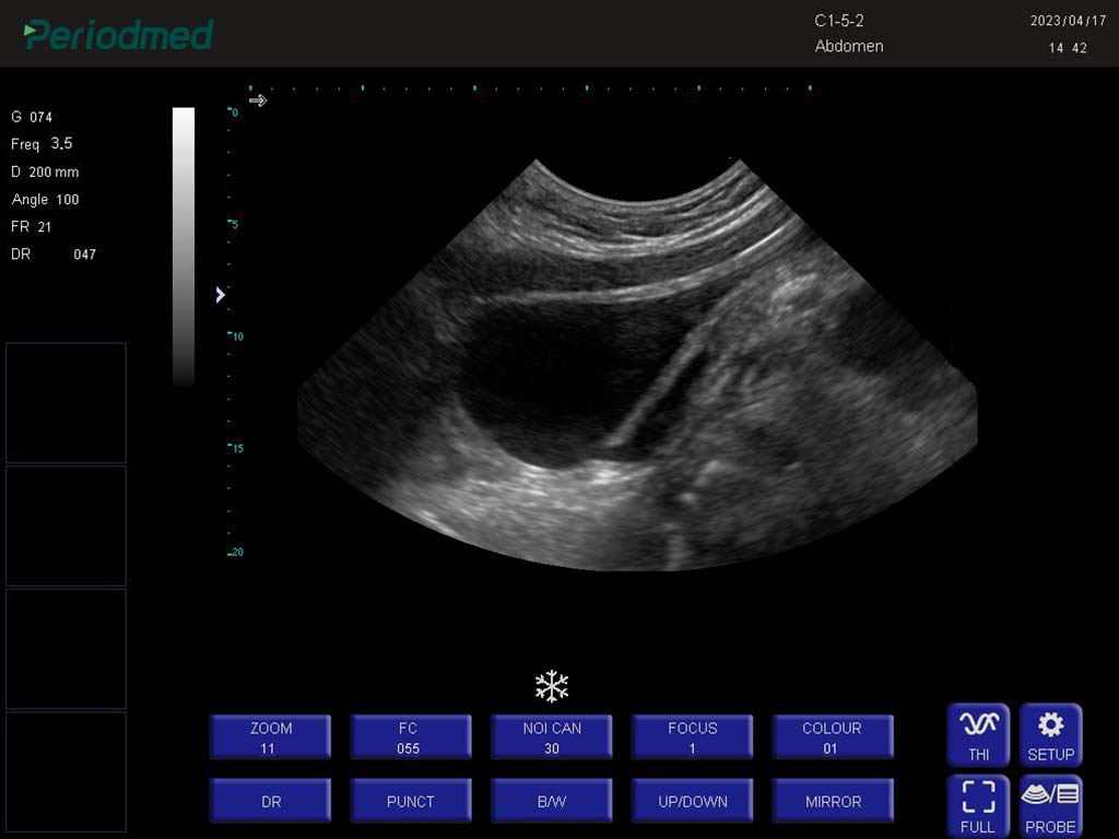
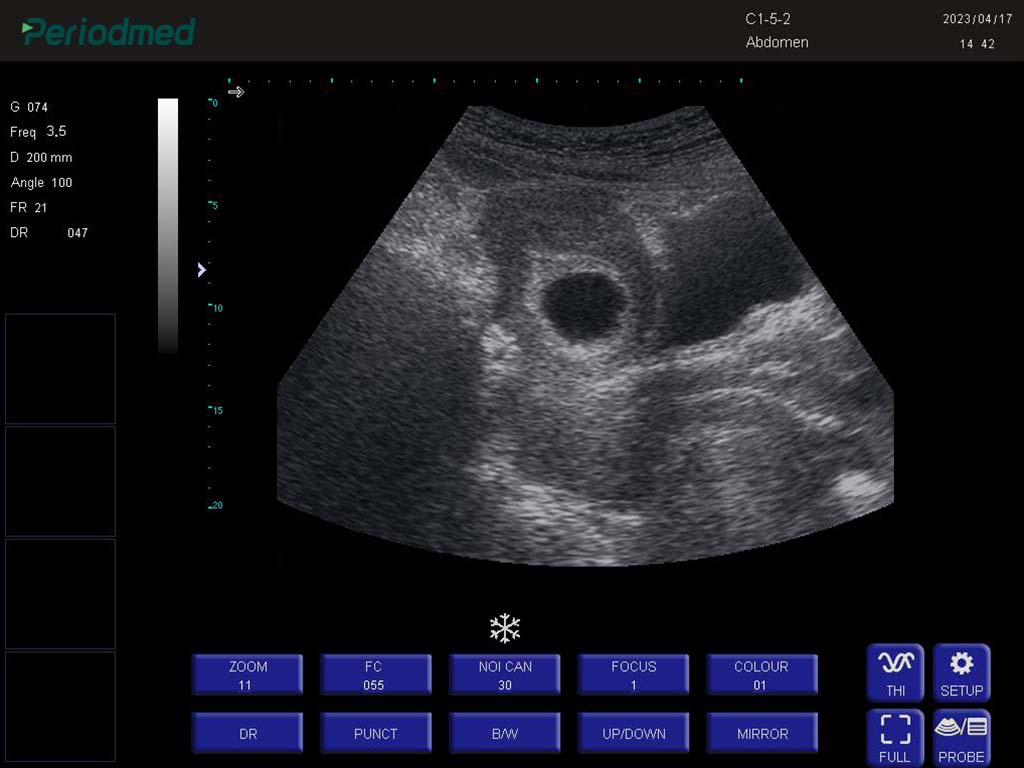
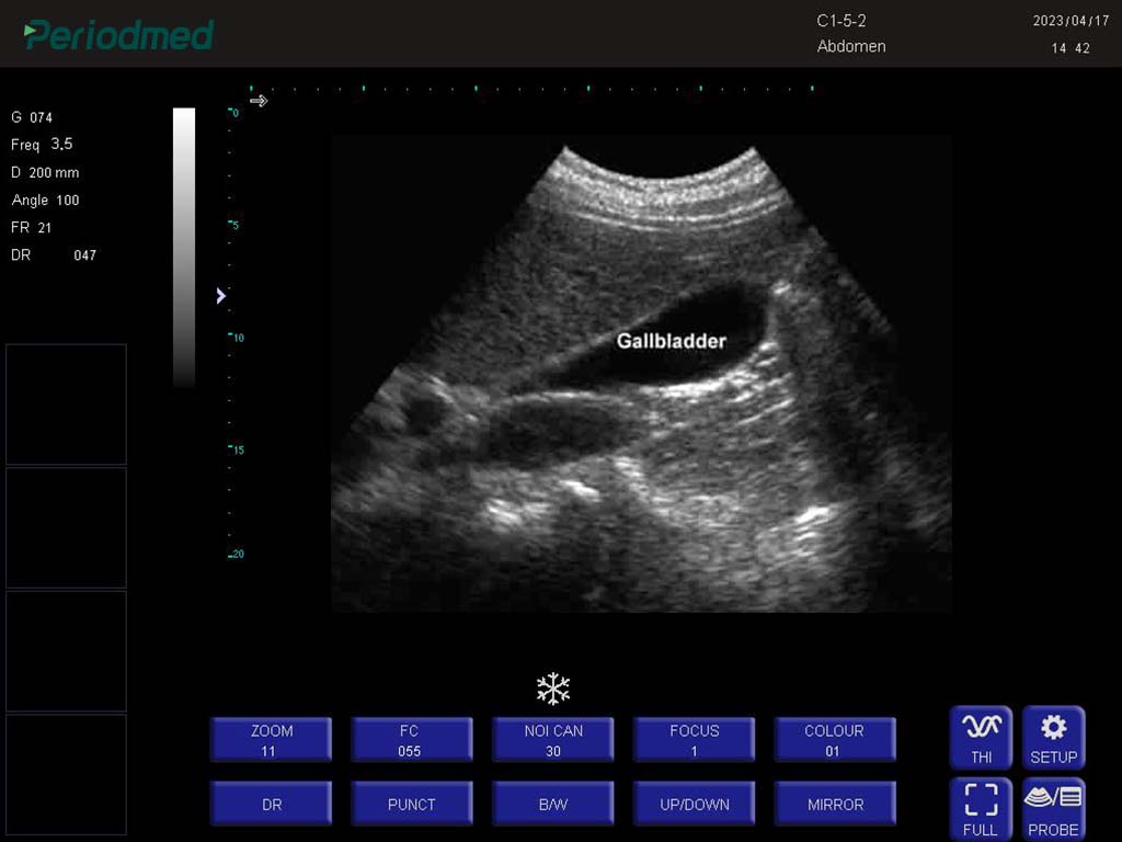
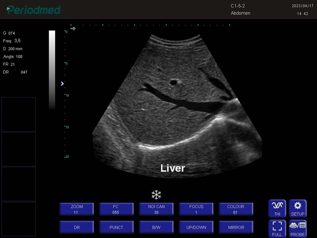
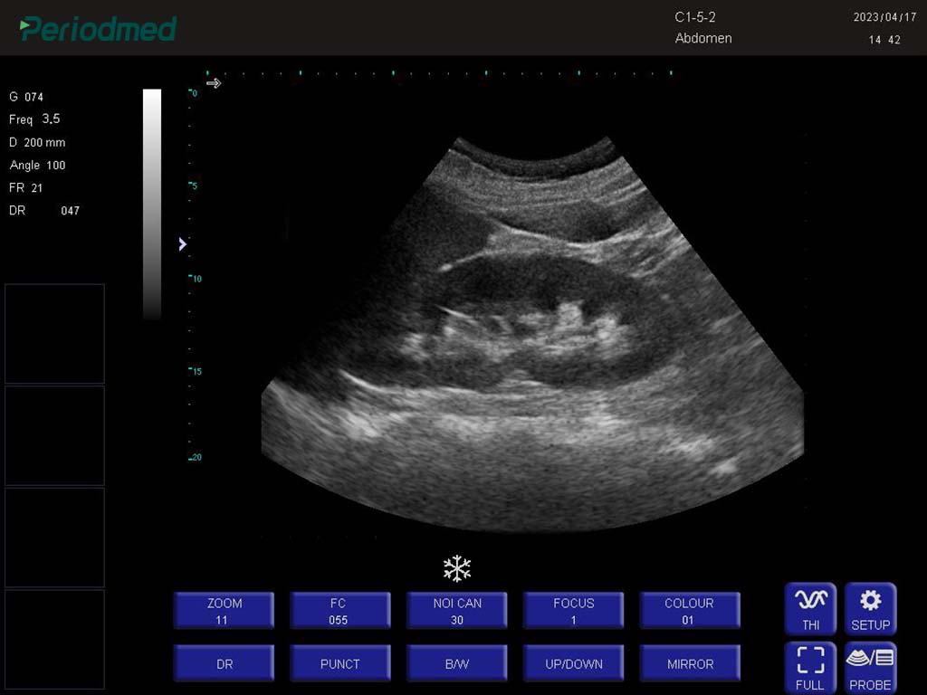
PU-DL121A is a desktop thyroid ultrasound machine with highly mobile, slim and smart design that permits easy transportation and storage. By combining advanced adaptive precise transmission and reception control with efficient multi-channel parallel processing technology, thyroid ultrasound machine features maximized computing speed for the best image performance.
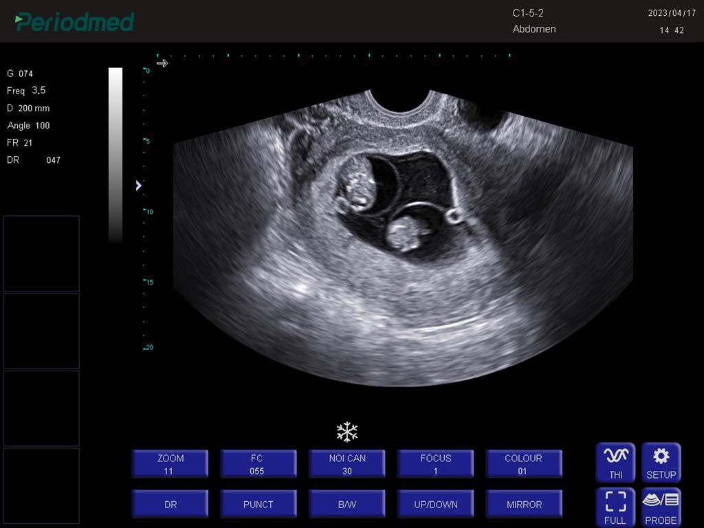
Built on extensive clinical experience, thyroid ultrasound machine allows doctors to characterize the thyroid gland, the surrounding tissues, and any nodules which may be present. Its high-frequency sound wave technology generates high-definition imagings, detecting various lesions, including thyroid nodules (solid or cystic, benign or malignant), goiter (enlargement of the thyroid), inflammations, cysts, and even abnormal lymph nodes in the neck.
PU-DL121A has various probes wiht a dedicated one-key optimization function that further simplifies thyroid ultrasound workflow, enabling real-time image enhancement, measurement, and calculation—all with a single press. It not only saves time but also ensures consistency in thyroid ultrasound image quality across different users and environments.
PU-DL121A uses harmonic Imaging Technology: using the second harmonics generated by the tissue boundary layer,THI significantly increase contrast resolution and improves thyroid ultrasound image quality. Imaging Technique: Automatic optimization, Noise cancellation, Speckle reduction imaging, Fine angle steer, Tissue harmonic imaging.
Thyroid ultrasound machine is equipped with a full set of standard accessories and optional expansion accessories. Both flexibility and practicality are ensured to meet both basic operational needs and personalized application scenarios. Standard accessories include USB, video cable, power cable, operator's manual, and optional accessories include printer, trolley, and metal box.
| Application | |||
| Abdomen | Obstetrics | Gynecology | Pediatrics |
| Vascular | Small Parts | Cardiology | Thyroid |
| MSK | Urology | ||
| Image Formats | |||
| AVI | BMP | JPG | PNG |
| DICOM | |||
| Transducer Types | |||
| Convex probe | Linear probe | Trans-rectal probe | Micro convex array |
| Trans-vaginal probe | |||
| Operating Mode | |||
| B | 2B | 4B | B/M |
| M | PW(Optional) | ||
| System Architecture | |||
| Physical Channels: 32 | Elements: 80/96/128 | THI:On/Off | 2 Probe connectors |
| Dynamic range:0-120db | Power:40-100 | 12 Inches High Resolution LED Monitor | Resolution:1024*768 |
| Triplex | |||
| B+CF | PDI | DPDI+PW | |
| Optional | |||
| High-frequency line array probe | Trolley | Printer | Metal box |


 EN
EN CN
CN