Tel/Whatsapp: +86 19352847010
Email: [email protected]

- Product
Ultrasound System
Life Information Monitoring System
Infusion & Syringe System
Solution
- About Us
- Media & Events
- Service
- Contact Us
Tel/Whatsapp: +86 19352847010
Email: [email protected]
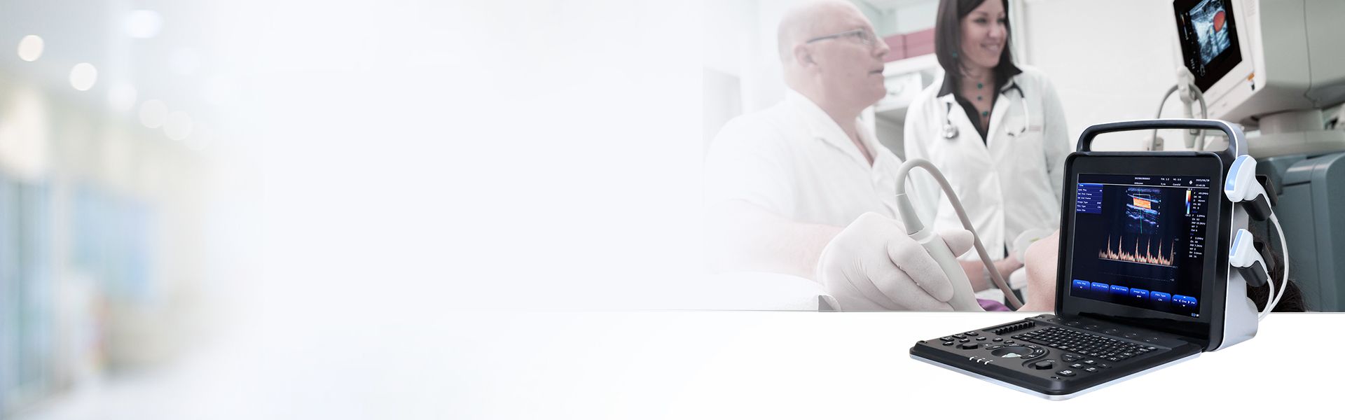
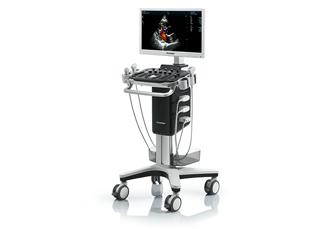
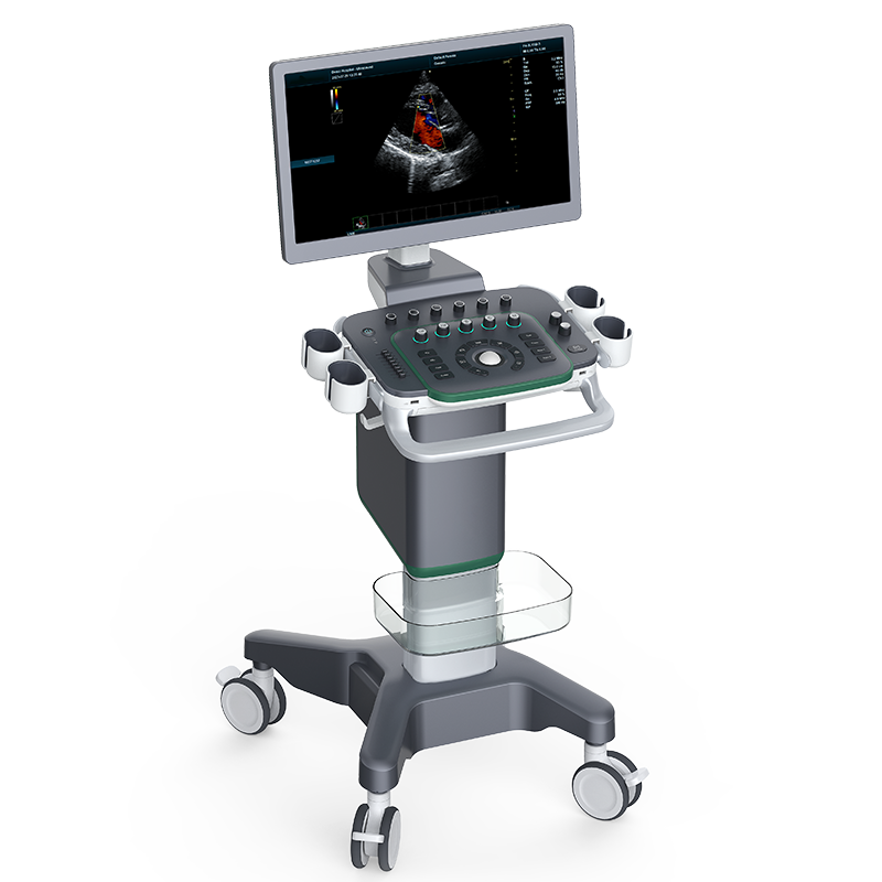
High-End Rehab Medicine POCUS | Revo T1

Revo T1 is a professional rehab medicine high-end POCUS system optimized for physiatry and neurological rehabilitation applications. Powered by multi-channel parallel processing and intelligent imaging algorithms, it delivers stable, high-definition soft tissue imaging. It supports comprehensive spasticity management ultrasound and muscle structure evaluation, serving as a core device for standardized rehabilitation clinical assessment.
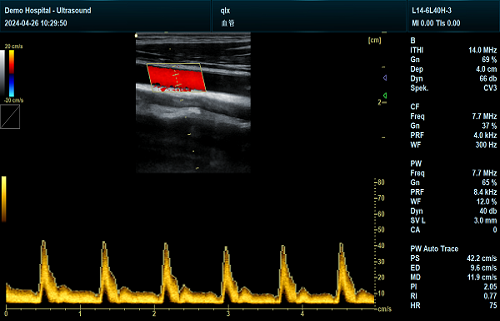
Integrating shear wave elastography muscle technology, Revo T1 enables accurate quantitative muscle ultrasound PMR assessment for spastic patients. It objectively measures muscle stiffness, elasticity and tissue texture, effectively differentiating spastic hypertonia from normal muscle tension. This quantitative data supports precise grading of muscular spasticity caused by neurological disorders for targeted rehabilitation planning.
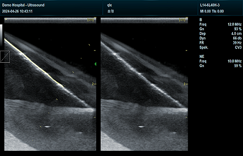
Featuring professional puncture enhancement and real-time dual imaging modes, Revo T1 ensures safe ultrasound-guided botulinum toxin injection spasticity procedures. It clearly visualizes target muscles, nerve branches and surrounding vessels, improving injection accuracy. It minimizes operational risks, optimizes spasticity relief effects, and standardizes minimally invasive intervention workflows in rehab clinics.
Equipped with optional 3D/4D imaging modes, the system supports precise 3D ultrasound muscle volume measurement rehab for atrophic and spastic muscles. It performs professional ultrasound muscle quality assessment neurological disorders, tracking muscle thickness, echo intensity and morphological changes. It provides reliable longitudinal data to monitor muscle degeneration and rehabilitation progress for neurological patients.
Revo T1 adopts iTouth one-click optimization, speckle reduction and spatial compound imaging to stabilize muscle scan quality. Built-in professional measurement packages and DICOM compatibility support standardized data recording. With multi-language adaptation and complete clinical modes, it fully meets high-precision muscle quantitative screening and long-term rehab follow-up in professional physiatry departments.
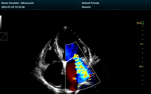
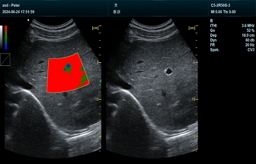
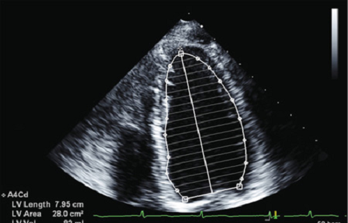
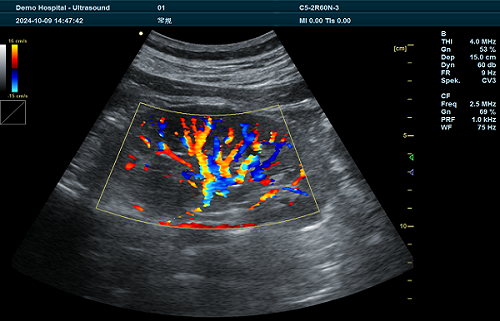
| Application | |||
| Generic | Obstetrics | Gynecology | Abdomen |
| Vascular | Small Parts | Urology | Thyroid |
| Cardiology | MSK | ||
| Image Formats | |||
| AVI | BMP | JPG | PNG |
| DICOM | |||
| Transducer Types | |||
| Convex probe | Linear probe | Phased array | Micro convex array |
| Volume array | Intracavity probe | ||
| Operating Mode | |||
| B | 2B | 4B | CW |
| M | CF | B/M | PW |
| PDI | Picture in Picture | Trapezoid | Panorama |
| 3D/4D(Optional) | Wide view(Optional) | Color M Mode | Anatomy M |
| TDI(Optional) | Needle Enhance Mode(Optional) | Contrast Enhance Mode(Optional) | |
| System Architecture | |||
| Elements: 64/128 | Frequency Range: 2.0-22.0 MHz | Channel:64 | Hard Drive:128G |
| Dynamic Range: ≥120db | 13.3 Inches touch screen | Resolution:1024*768 | Beam forming 4 |
| Memory:4G | 21 Inches High Resolution LED Monitor | ||
| Triplex | |||
| B+CF | PDI | DPDI+PW | |
| Languages | |||
| English | Arabic | French | Indonesian |
| Italian | Portuguese | Spanish | Chinese |
| German | Russian | ||


 EN
 CN