EN
EN CN
CN
The Future of Ultrasound Technology: Innovations and Breakthroughs
Ultrasound technology has undergone remarkable advancements in recent years, revolutionizing diagnostic imaging and medical applications. As non-invasive imaging continues to gain prominence, researchers and engineers are pushing the boundaries of what ultrasound can achieve. From artificial intelligence integration to high-resolution imaging and portable devices, ultrasound technology is evolving to enhance diagnostic accuracy, efficiency, and accessibility.
AI-Powered Ultrasound: Enhancing Diagnostic Precision
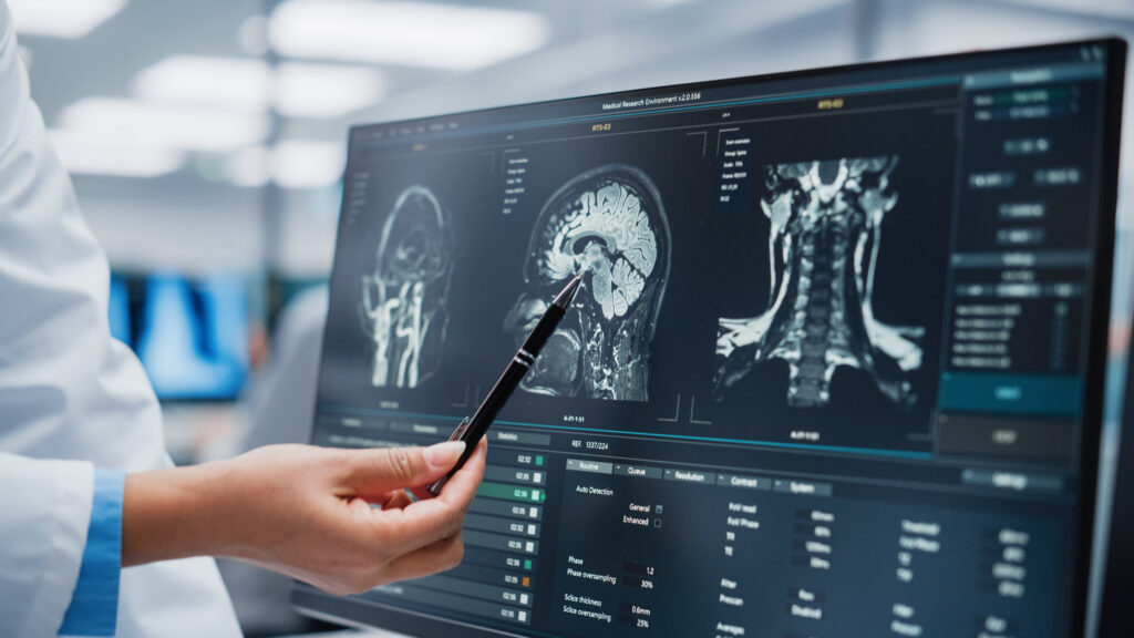
Artificial intelligence (AI) is playing a pivotal role in transforming ultrasound imaging. Traditional ultrasound scans rely heavily on the expertise of sonographers and radiologists, making interpretation subjective. However, AI-powered algorithms are now being integrated into ultrasound systems to assist with real-time image analysis, reducing operator dependency and minimizing errors.
Deep learning models can automatically identify abnormalities, differentiate between benign and malignant tumors, and provide quantitative assessments in obstetrics, cardiology, and oncology. AI-powered ultrasound has demonstrated potential in early disease detection, such as liver fibrosis, breast cancer, and thyroid nodules, by analyzing subtle patterns that may be overlooked by human interpretation. As AI continues to evolve, its ability to learn from vast datasets will significantly enhance diagnostic capabilities, making ultrasound a more reliable and efficient tool.
High-Resolution and 3D/4D Imaging
One of the major challenges of conventional ultrasound has been its limited image resolution compared to other imaging modalities like MRI and CT scans. However, recent innovations in high-frequency transducers and advanced signal processing have led to significant improvements in image clarity.
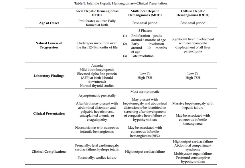
High-frequency ultrasound (HFU) enables detailed visualization of superficial structures such as tendons, nerves, and skin layers. Meanwhile, 3D and 4D ultrasound imaging have gained popularity, particularly in obstetrics and gynecology. 3D imaging constructs volumetric representations of organs, while 4D imaging adds the dimension of real-time movement, allowing clinicians to observe fetal movements, heart valve motions, and blood flow dynamics.
In cardiology, 4D echocardiography provides comprehensive views of the heart, helping physicians assess complex conditions like valve regurgitation and congenital heart defects with greater accuracy. With continued advancements, 3D and 4D ultrasound are expected to replace traditional 2D imaging in many medical applications.
Miniaturization and Wearable Ultrasound Devices
The trend toward miniaturization has led to the development of portable and even wearable ultrasound devices. Traditional ultrasound machines are often large, expensive, and confined to hospital settings, limiting access in remote or underdeveloped regions.
Handheld ultrasound devices, such as those connected to smartphones and tablets, are becoming more common in point-of-care applications. These devices provide emergency physicians, paramedics, and primary care doctors with real-time imaging capabilities outside traditional hospital environments.
Wearable ultrasound patches, a recent breakthrough, have been designed to continuously monitor vital organs such as the heart and lungs. These soft, flexible devices adhere to the skin and provide long-term ultrasound imaging without requiring a skilled operator. They hold great potential for monitoring chronic diseases, assessing sports injuries, and even detecting early signs of conditions like deep vein thrombosis (DVT) or internal bleeding.
Therapeutic Ultrasound: Beyond Imaging
While ultrasound is widely recognized for its diagnostic applications, it is also making strides in therapeutic fields. High-Intensity Focused Ultrasound (HIFU) is a non-invasive treatment method that uses ultrasound waves to destroy abnormal tissue, such as tumors, without surgery or radiation.
HIFU has shown promising results in treating prostate cancer, uterine fibroids, and certain neurological disorders like essential tremors and Parkinson’s disease. By precisely targeting affected tissues while sparing surrounding structures, HIFU minimizes side effects compared to traditional treatments.
Another emerging application is ultrasound-assisted drug delivery. Microbubbles injected into the bloodstream can be manipulated using ultrasound waves to temporarily open the blood-brain barrier (BBB), allowing medications to reach the brain more effectively. This technique holds significant promise for treating neurological conditions like Alzheimer's and brain tumors, which have been challenging due to the BBB’s natural defense mechanisms.
Contrast-Enhanced Ultrasound (CEUS) for Improved Vascular Imaging
Traditional ultrasound imaging has limitations in visualizing blood flow and vascular structures, often requiring supplementary imaging techniques like Doppler ultrasound or angiography. However, Contrast-Enhanced Ultrasound (CEUS) is changing the landscape of vascular imaging.
CEUS involves injecting microbubble contrast agents into the bloodstream, which improve ultrasound signal reflections and enhance image clarity. This technique enables real-time assessment of organ perfusion, tumor vascularity, and cardiovascular conditions. Unlike CT and MRI contrast agents, CEUS contrast materials have a lower risk of kidney toxicity, making them a safer option for patients with renal impairment. Learn more about Periodmed products such as patient monitors, and ultrasound for animals.