Tel/Whatsapp: +86 15005204265
Email: [email protected]
- Home
- Product
Ultrasound System
Life Information Monitoring System
Infusion & Syringe System
Solution
- About
- Media & Events
- Service & Support
- Contact Us
Tel/Whatsapp: +86 15005204265
Email: [email protected]
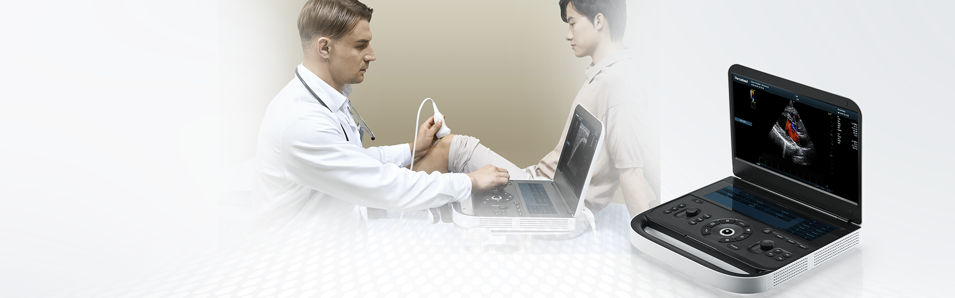

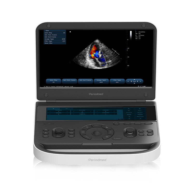
Ultra-Portable and Highly Flexible | Revo 9

Revo 9 heart ultrasound system is conceived to optimize heart ultrasound examination workflow, making it a really productive platform. Our heart ultrasound device uses sound waves to create real-time images of patient's heart, allowing doctors to assess its structure, function, and blood flow through the heart. The ultrasound procedure helps doctors assess heart conditions like heart failure, valvular regurgitation, and heart murmurs.
Designed with smart ergonomics to be light and silent, Revo 9 brings comfort to patients and operators when doing heart ultrasound(echocardiogram) using advanced technologies provided by Periodmed which maximizes productivity. The non-invasive ultrasound test uses sound waves to create moving images of the heart, allowing doctors to assess its structure, function, and blood flow through its chambers and valves.
Periodmed ProOpti™ system utilizes automatic structure detection algorithms to enhance heart ultrasound image quality by sharpening edges and ensuring smooth, uniform tissue representation. This innovative feature enhances heart ultrasound diagnostic accuracy by providing clearer and more detailed images. PanoramaVision™ technology captures a series of two-dimensional sectional images, which are reconstructed into an ultra-wide sectional image with a continuous field of view.
By employing PrismX™ spatial composite imaging technology, our heart ultrasound system enhances contrast, fine resolution, and spatial resolution. PureSight™ technology reduces speckle artifacts, improves smoothness along preferred edge directions, and maintains consistent local average gray levels. This results in clearer and more visually appealing images, enhancing heart ultrasound diagnostic confidence.
Revo 9 is equipped with a full set of standard accessories and optional expansion accessories. Both flexibility and practicality are ensured to meet both basic operational needs and personalized application scenarios. Standard accessories include USB, video cable, power cable, operator's manual, and optional accessories include printer, trolley, and metal box.


| Application | |||
| Generic | Obstetrics | Gynecology | Abdomen |
| Vascular | Small Parts | Urology | Thyroid |
| Cardiology | MSK | ||
| Image Formats | |||
| AVI | BMP | JPG | PNG |
| DICOM | |||
| Transducer Types | |||
| Convex probe | Linear probe | Phased array | Micro convex array |
| Volume array | Intracavity probe | ||
| Standard Mode | |||
| B | 2B | 4B | CW |
| M | CF | PDI | PW |
| DPDI | Picture in Picture | Trapezoid | Panorama |
| Color M Mode | Anatomy M | TDI(Optional) | Wide View(Optional) |
| 3D/4D Mode(Optional) | Needle Enhance Mode(Optional) | Contrast Enhance Mode(Optional) | SMV(Optional) |
| Elastography(Optional) | |||
| System Architecture | |||
| Elements: 64/128/160/192 | Frequency Range: 2.0-22.0 MHz | Channel:64 | Hard Drive:256G |
| Dynamic Range: 36-120db | 13.3 Inches touch screen | Resolution:1920*1080 | Beam forming 16 |
| Memory:8G | 23.8 Inches High Resolution LED Monitor | ||
| Languages | |||
| English | Arabic | French | Indonesian |
| Italian | Portuguese | Spanish | Chinese |
| German | Russian | ||


 EN
EN CN
CN