Tel/Whatsapp: +86 19352847010
Email: [email protected]

- Home
- Product
Ultrasound System
Life Information Monitoring System
Infusion & Syringe System
Solution
- About
- Media & Events
- Service & Support
- Contact Us
Tel/Whatsapp: +86 19352847010
Email: [email protected]
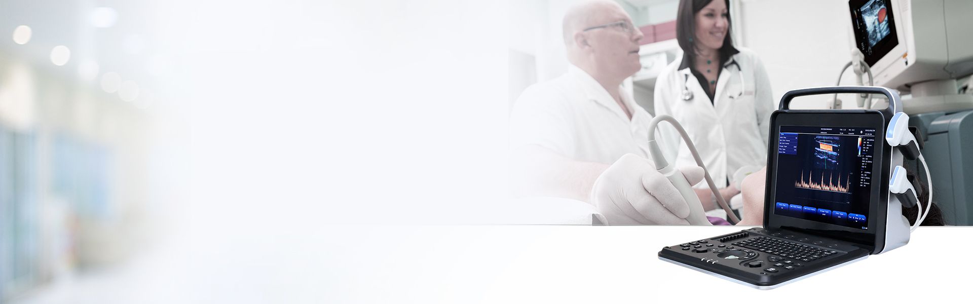
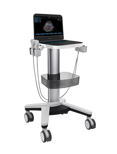
Cardiac Anomaly Screening | Revo 9

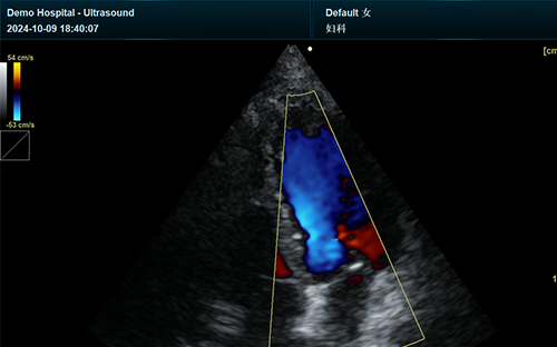
Revo 9 advanced system delivers professional structural heart ultrasound for standardized cardiac structural and valvular assessment. Equipped with 192-element imaging channels and optimized cardiac modes, it captures fine anatomical details of valves, myocardium and pericardium. It serves as a reliable POCUS for valvular heart disease screening, supporting early detection of congenital and degenerative structural cardiac lesions in clinical routines.
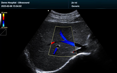
Featuring high-sensitive CW Doppler handheld structural heart functionality, Revo 9 enables precise portable ultrasound aortic valve gradient measurement. Its wide dynamic range and intelligent noise reduction eliminate blood flow artifacts, ensuring accurate peak and mean transvalvular pressure gradient data. This quantitative evaluation lays objective foundation for valvular stenosis and regurgitation severity grading.
Specialized for TAVR guidance POCUS workflows, Revo 9 provides real-time handheld echo visualization for transcatheter aortic valve replacement procedures. Multi-angle biplane imaging clearly displays aortic root structure and valve positioning. It assists clinicians in preoperative sizing, intraoperative prosthesis alignment and immediate post-procedural structural assessment to reduce procedural risks.
Supporting precise pericardiocentesis ultrasound guidance, the system delivers clear real-time imaging of pericardial effusion and adjacent vital structures. Integrated needle enhancement mode improves puncture accuracy and avoids myocardial or vascular injury. As core cardiac ultrasound interventional guidance equipment, it ensures safe, efficient emergency pericardial drainage in critical cardiac scenarios.
Revo 9 integrates comprehensive cardiac imaging modes including TDI, CW/PW Doppler and panoramic scanning. Advanced PrismX™ and PureSight™ technologies stabilize image quality for complex cardiac assessments. With DICOM compatibility and professional cardiac calculation packages, it meets full-cycle demands for screening, intervention guidance and follow-up of structural and valvular heart diseases.
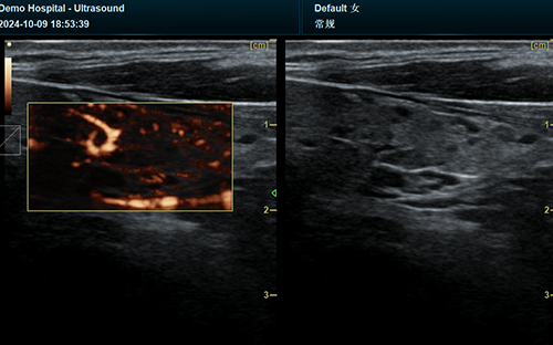
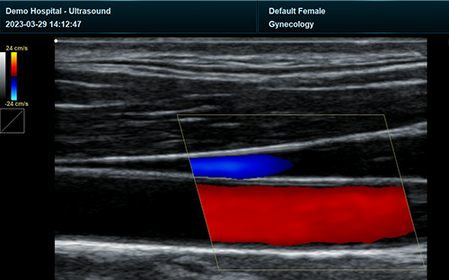
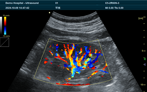
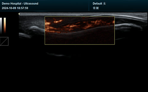
| Application | |||
| Generic | Obstetrics | Gynecology | Abdomen |
| Vascular | Small Parts | Urology | Thyroid |
| Cardiology | MSK | ||
| Image Formats | |||
| AVI | BMP | JPG | PNG |
| DICOM | |||
| Transducer Types | |||
| Convex probe | Linear probe | Phased array | Micro convex array |
| Volume array | Intracavity probe | ||
| Standard Mode | |||
| B | 2B | 4B | CW |
| M | CF | PDI | PW |
| DPDI | Picture in Picture | Trapezoid | Panorama |
| Color M Mode | Anatomy M | TDI(Optional) | Wide View(Optional) |
| 3D/4D Mode(Optional) | Needle Enhance Mode(Optional) | Contrast Enhance Mode(Optional) | SMV(Optional) |
| Elastography(Optional) | |||
| System Architecture | |||
| Elements: 64/128/160/192 | Frequency Range: 2.0-22.0 MHz | Channel:64 | Hard Drive:256G |
| Dynamic Range: 36-120db | 13.3 Inches touch screen | Resolution:1920*1080 | Beam forming 16 |
| Memory:8G | 23.8 Inches High Resolution LED Monitor | ||
| Languages | |||
| English | Arabic | French | Indonesian |
| Italian | Portuguese | Spanish | Chinese |
| German | Russian | ||


 EN
EN CN
CN