Tel/Whatsapp: +86 19352847010
Email: [email protected]

- Home
- Product
Ultrasound System
Life Information Monitoring System
Infusion & Syringe System
Solution
- About
- Media & Events
- Service & Support
- Contact Us
Tel/Whatsapp: +86 19352847010
Email: [email protected]
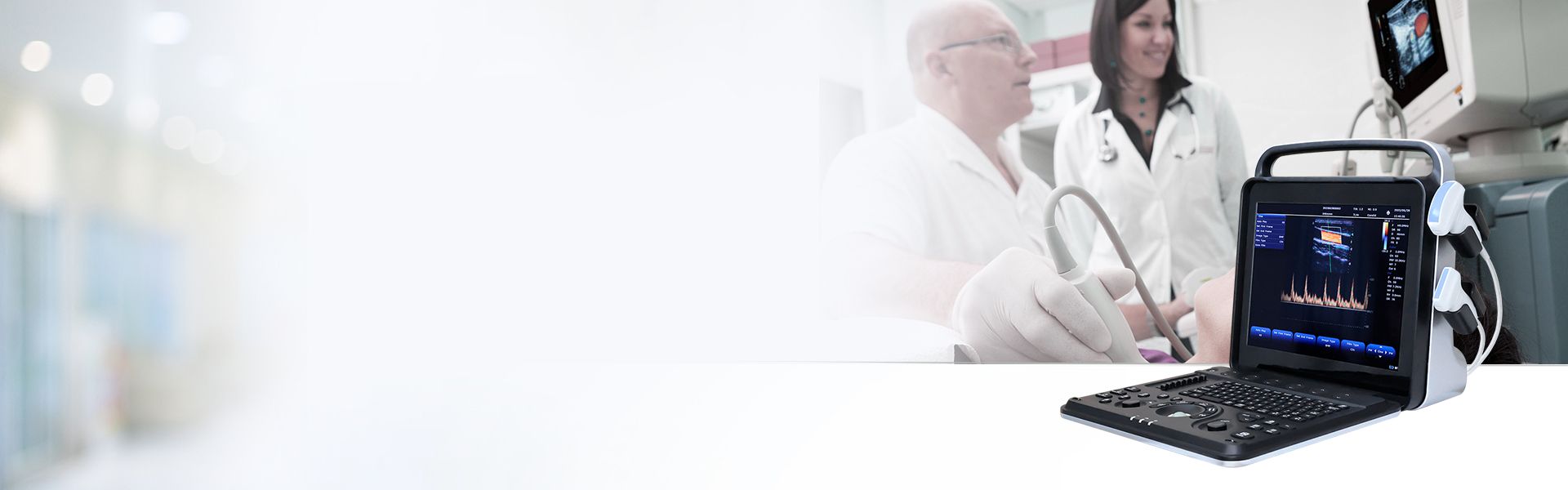
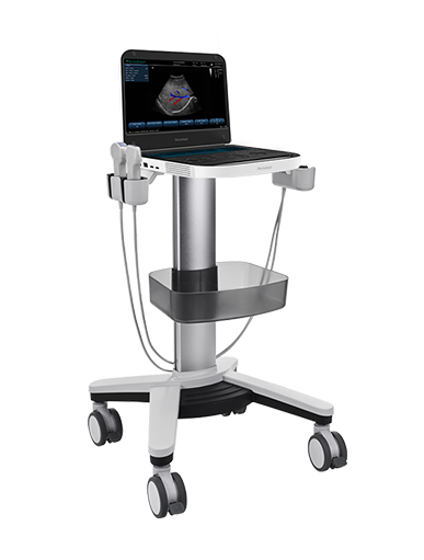
Bedside Quantitative Echo Assessment | Revo 9

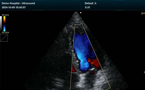
Revo 9 professional system is engineered for reliable cardiac ultrasound outpatient examinations, delivering standardized bedside echo quantitative assessment in clinic settings. With advanced multi-channel beamforming and image optimization algorithms, it produces clear cardiac structural and hemodynamic images. It enables fast, repeatable outpatient POCUS screening for routine cardiac function and lesion evaluation.
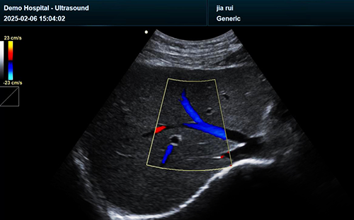
Supporting accurate POCUS ejection fraction quantification, Revo 9 delivers stable handheld ultrasound LVEF measurement clinic results for daily cardiology workflows. High-resolution B-mode imaging clearly presents ventricular wall motion and chamber geometry. Clinicians obtain objective left ventricular systolic function data, supporting early heart failure screening and consistent outpatient condition grading.
Equipped with sensitive tissue Doppler imaging, the device supports professional TDI e' assessment handheld to evaluate myocardial diastolic performance. It captures subtle tissue velocity changes unavailable in conventional scanning. These valuable diastolic parameters provide strong prognostic evidence, helping clinicians differentiate early diastolic dysfunction and stratify long-term cardiac risk in outpatient patients.
As a powerful Doppler echocardiography handheld solution, Revo 9 enables reliable PW Doppler cardiac output outpatient calculation. It accurately acquires LVOT-VTI and hemodynamic velocity data to evaluate real-time cardiac output and fluid status. This handheld cardiac Doppler workflow standardizes outpatient hemodynamic quantification for precise and evidence-based cardiac treatment adjustments.
Featuring PureSight™ and PrismX™ imaging enhancement, Revo 9 minimizes artifacts during continuous outpatient scanning. Integrated automatic measurement packages streamline quantitative analysis of LVEF, e’ and VTI indicators. Compatible with DICOM storage and multi-mode cardiac scanning, it supports complete follow-up recording and prognostic tracking for chronic cardiac outpatient diseases.
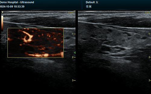
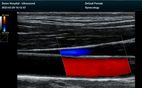
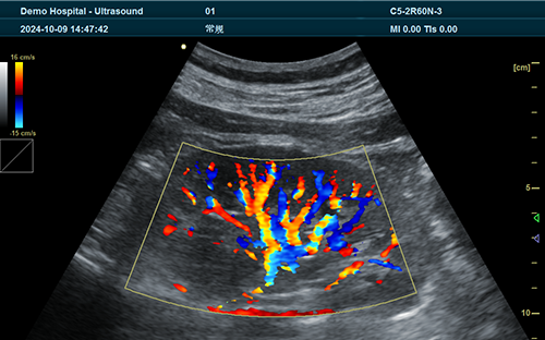
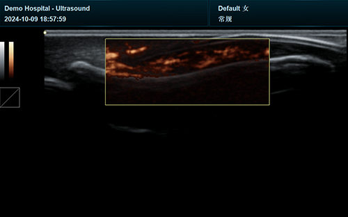
| Application | |||
| Generic | Obstetrics | Gynecology | Abdomen |
| Vascular | Small Parts | Urology | Thyroid |
| Cardiology | MSK | ||
| Image Formats | |||
| AVI | BMP | JPG | PNG |
| DICOM | |||
| Transducer Types | |||
| Convex probe | Linear probe | Phased array | Micro convex array |
| Volume array | Intracavity probe | ||
| Standard Mode | |||
| B | 2B | 4B | CW |
| M | CF | PDI | PW |
| DPDI | Picture in Picture | Trapezoid | Panorama |
| Color M Mode | Anatomy M | TDI(Optional) | Wide View(Optional) |
| 3D/4D Mode(Optional) | Needle Enhance Mode(Optional) | Contrast Enhance Mode(Optional) | SMV(Optional) |
| Elastography(Optional) | |||
| System Architecture | |||
| Elements: 64/128/160/192 | Frequency Range: 2.0-22.0 MHz | Channel:64 | Hard Drive:256G |
| Dynamic Range: 36-120db | 13.3 Inches touch screen | Resolution:1920*1080 | Beam forming 16 |
| Memory:8G | 23.8 Inches High Resolution LED Monitor | ||
| Languages | |||
| English | Arabic | French | Indonesian |
| Italian | Portuguese | Spanish | Chinese |
| German | Russian | ||


 EN
EN CN
CN